Introducing ScopeCare Service Agreements from DB Surgical

ScopeCare Service Agreements from DB Surgical – The Local Experts in Surgical Microscopes
For Maximum Performance, Superior Reliability, and Budget Peace of Mind


The ScopeCare program combines exceptional service with fixed repair costs, to ensure your investment is protected and that you’ll have the power to shape the future of better outcomes. To learn more, watch the video presentation below or download the brochure here: ScopeCare Service Agreement Brochure (~700KB PDF file)


ScopeCare plans are tailored to your needs with three different agreement options, which are shown in the chart on the brochure. All ScopeCare plans come with two preventative maintenance visits per year, our rapid response guarantee, unlimited technical support, and unlimited software updates. Additionally:

  • The Prevent plan provides a discount of 10% on labor, travel, and mileage.
  • The Plus plan provides a discount of 50% on labor, travel, and mileage along with a 5% discount on replacement parts.
  • The Premium plan provides a discount of 100% on labor, travel, and mileage along with a 10% discount on replacement parts.

ScopeCare Service Agreements are designed to provide maximum performance, superior reliability, and budget peace of mind. Guard against unplanned downtime that disrupts patient care, with: preventative maintenance visits, priority service calls, and guaranteed response times all from the most highly qualified technicians serving the Southeast. Depend on DB Surgical for help with support from the largest local service team of: Leica-certified technicians who are reachable directly via phone, text, or email and that have proprietary knowledge and access to parts for accurate diagnosis and resolution. Facilitate budget planning and avoid costly repairs through: multi-year service agreements with fixed pricing for predictable maintenance costs; all while working with the only team that can maintain warranties and repair to original guidelines.


ScopeCare Service Agreements from DB Surgical – The Local Experts in Surgical Microscopes
For Maximum Performance, Superior Reliability, and Budget Peace of Mind

To protect your surgical microscope, contact us today for your customized proposal

Augment Your Reality: Introducing the ARveo Digital Augmented Reality Microscope

A Single Vision for the Future with Augmented Reality

First, came the microscope, which transformed previously unseen parts of the body into entire fields of expertise. Then came surgical fluorescence, which helped reveal more detail within the body. Now, augmented reality built directly into the ARveo digital surgical microscope is set to push the boundaries of surgical visualization, by making the previously unknowable, known.


Discover the benefits of the ARveo


The ARveo digital augmented reality microscope with groundbreaking GLOW AR fluorescence technology empowers surgical decision-making by providing:

  • Augmented digital imaging for precise, real-time information
  • The freedom to choose what you see and how you see it
  • Flexible options to share your view in the OR and beyond

Professor Guzman talks about the future of neurosurgical imaging


In this video Professor Raphael Guzman, MD talks about the benefit of having more information during surgery by integrating augmented imaging technologies, such as GLOW800 AR fluorescence and neuronavigation, in one microscope.


Advantages of GLOW800 AR fluorescence compared to NIR imaging


Compare visualization of cerebrovascular flow with GLOW800 AR fluorescence to traditional near infrared (NIR) imaging in this video. See how GLOW800 augmented reality (AR) fluorescence takes the high contrast of NIR imaging in combination with ICG and fuses it with the naturally-colored microscope view in real-time


The ARveo is our most advanced microscopy imaging solution to date. Designed for the most complex surgical interventions, it features sophisticated digital imaging technology, including imaging sensors and advanced alogorithms, outstanding optics and the ability to capture and share surgery in 3D. What’s more, only ARveo with GLOW AR technology provides a real-time augmented reality view of the surgical field, supporting you to assess and perform procedures with complete confidence.


Contact DB Surgical for more information.

Republished from Leica Microsystems.

Leica Microsystems Featured On “Innovations with Ed Begley Jr”

Leica Microsystems is advancing surgical microscopy by integrating enhanced digital technologies directly into the operating microscope. See the latest advances in surgical microscopy, and learn how Leica Microsystems is providing surgeons with better insight to improve their decision-making at the point of care.

Four prominent surgeons and Leica Microsystems President Markus Lusser discuss the many ways Leica Microsystems is pushing the boundaries of visualization in the operating environment by integrating digital technologies directly into the surgical microscope.

  • Seenu M. Hariprasad, MD
    Shui-Chin Lee Professor of Ophthalmology and Visual Science
    Chief, Vitreoretinal Service
    The University of Chicago Medicine, USA
  • James Reidy, MD
    Assoc. Professor of Ophthalmology & Visual Science
    Vice Chair for Clinical Operations
    Medical Director of the Ophthalmology Outpatient Center
    The University of Chicago Medicine, USA
  • Cleopatra Charalampaki, MD, PhD
    Professor of Neurosurgery, Department of Neurosurgery
    Cologne Medical Center, Germany
  • Markus Lusser
    President Leica Microsystems
  • Makoto Nakamura, MD, PhD
    Professor of Neurosurgery, Director of Neurosurgery
    Cologne Medical Center, Germany

Contact DB Surgical for more information.

Republished from Leica Microsystems.

Leica FL560 Achieves First FDA Clearance for Cerebrovascular Fluorescence with Fluorescein

Leica Microsystems is the first company in the U.S. to receive FDA 510(k) clearance for the visualization of cerebrovascular blood flow in conjunction with fluorescein.


WETZLAR, Germany, Aug. 29, 2017 /PRNewswire/ — Leica Microsystems is proud to be the first company to attain FDA 510(k) clearance for its FL560 fluorescence microscope filter for visualization of cerebrovascular blood flow in conjunction with the dye fluorescein. When integrated into a M530 OH6 neurosurgical microscope, FL560 fluorescence provides real-time, high-contrast visualization of both cerebral anatomy in native color and fluorescent blood flow. With this combined view, the surgeon has more information to aid assessment and decision-making during vascular neurosurgery.


Assessing cerebral anatomy and vascular flow, particularly in smaller vessels and the areas they perfuse, can be challenging under white light or with traditional near-infrared which only provides a black and white image. By integrating the FL560 fluorescence filter into the M530 OH6 neurosurgical microscope, the surgeon is able to view anatomical structures in white-light and fluorescent blood flow simultaneously in the oculars. This is achieved by combining premium microscope optics and illumination with a sophisticated, proprietary fluorescence filter design that effectively separates fluorescence excitation light and the observation spectrum. The result is a high-contrast image where anatomy is clearly visible and even the smallest vessels delineated.

Download the U.S. brochure for FL560 here.

“Leica Microsystems has been a leader in advanced surgical visualization for decades. We have achieved a number of ‘firsts’ over the years including the first FDA 510(k) clearance of a microscope for intra-operative angiography (FL800) and the first company offering a surgical microscope to integrate three types of fluorescence capabilities (TriFluoro). I am proud that our team again was able to be the innovation leader by being the first company to achieve FDA clearance for cerebrovascular fluorescence imaging with fluorescein (FL560). This demonstrates our commitment to deliver clinically relevant and validated solutions to clinicians that allow them to make crucial surgical decisions and as a result improve patient outcomes,” says Markus Lusser, President Leica Microsystems.

FL560 fluorescence can aid the surgeon’s visualization and thus surgical decisions in a variety of neurovascular cases including Arteriovenous Malformations (AVM) and aneurysms.

FL560 fluorescence from Leica Microsystems has been designed to allow full integration into existing or new M530 OH6 neurosurgical microscopes at any time. The M530 OH6 microscope was the first to feature TriFluoro technology. This enables FL560 and two other types of fluorescence to be installed into a single microscope. Full integration also facilitates a smooth workflow with one-touch activation of different fluorescence modes via microscope handgrip or wireless footswitch, further supporting the surgical workflow.


Contact DB Surgical for more information.

Republished from PR Newswire and Leica Microsystems. 

Brain Aneurysm Clipping with the Leica M530 OH6 Neurosurgical Microscope

Recording of a brain aneurysm clipping by Vik Udani, MD, Division of Neurosurgery, Senta Clinic, San Diego, CA.


Patient is a 38-year-old female with a history of hypertension and migraines. She was found to have an 8mm unruptured right middle cerebral artery aneurysm and underwent a right frontal temporal craniotomomy for aneurysm clipping.

Dr. Udani on the M530 OH6 surgical microscope: “One of my favorite aspects of the OH6 is the amount of illumination on the surgical field. The light transmission provides sharp, crisp, bright images of fine vascular structures, which greatly enhances my visualization throughout the entire procedure. The improved illumination as well as the integrated camera also provides greater clarity when using the ICG activated filter giving me greater confidence in my surgical decisions.”

Watch this video and experience for yourself: Bright 400-Watt Xenon light system for concentrated light beam into deep cavities; high resolution and depth of focus with exclusive FusionOptics technology.

Find out more about the M530 OH6 premium surgical microscope for neurosurgery: Leica M530 OH6 with FusionOptics

Case and video provided courtesy of Vik Udani, MD.


Contact DB Surgical for More Information.

Republished from Leica Microsystems.

Navigating Through the Brain

Image Guided Surgery with microscope image injection aids visualization and orientation during neurosurgery


Image Guided Surgery
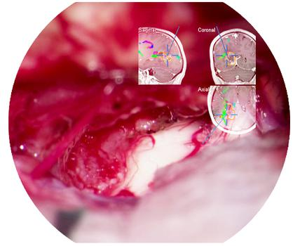
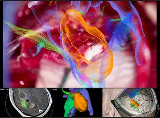
One of the challenges of neurosurgery is orientation at the surgical site. When resecting tumors, removing arteriovenous malformations, or clipping aneurysms, surgeons often have to work near healthy and functional brain tissue. When resecting the tumor, the challenge is always to spare as much healthy tissue as possible. Neuronavigation technology, also referred to as Image Guided Surgery (IGS) enables surgeons to plan the ideal approach before making a cut and helps to execute that plan by providing intraoperative orientation.

At the base of the human skull there is a particularly high concentration of vital structures: blood vessels, nerves, centers for hormonal control of bodily functions, and centers for regulating breathing, blood pressure, and heart activity. Brain tissue cannot regenerate itself, so even the smallest injury can cause irreversible, profound brain damage, leaving patients with a permanent disability. This also makes the treatment of disease very difficult. Malignant gliomas, which account for 45% to 50% of all brain tumors, infiltrate into brain tissue as they grow. Even under a surgical microscope, peripheral tumor regions are hard to differentiate from healthy tissue.

Continuous real-time view of the surgical field

Neuronavigation works like a GPS system, enabling accurate position tracking of instruments within the patient’s anatomy. Surgeons can outline the tumor, identify organs at risk, and define the optimal access pathway to the tumor to prevent injury to healthy structures.

Before surgery, CT or MR images of the patient are taken. In the operating room, the navigation system matches these images up with the live patient images using a wireless laser pointer. The pointer identifies surface points on the patient’s skin which are transmitted to the navigation system via an infrared camera. During the procedure, the camera also transmits the locations of the instruments and equipment – including the microscope – which are outfitted with reflective markers.

The patient images prepared prior to surgery are loaded into the navigation system and reconstructed into images on the monitor or injected into the oculars of the surgical microscope. This gives the surgeon a continuous, real-time map of the position of the instruments in relation to the brain structures and the tumor.

Image injection for a complete picture

To view the neuronavigation data, surgeons normally need to raise their heads from the microscope to look at the navigation screen. Thanks to image injection systems, like the new CaptiView from Leica, and in combination with the new picture-in-picture feature from Brainlab, the surgeon can see navigation data directly in the microscope’s eyepieces. The full-HD display of CaptiView image injection with LED backlighting provides bright, crisp injected images with distinct margins. Neuronavigation data from the Image Guided Surgery system can be viewed either in the right, the left, or both eyes and the CaptiView system is also able to display real-time HD images from an endoscope and FL800 vascular fluorescence. ”By having all information directly in the microscope’s ocular, the surgeon does not have to reorient between the microscope and the navigation screen, but can remain fully concentrated on the patient during the whole procedure”, explains David A Smith, Product Manager at Leica Microsystems. Furthermore, injected images are also displayed in the assistant binoculars and can be recorded in full HD quality, supporting teaching both inside and outside of the OR.

Microscope integration offers new imaging options

Brainlab is one of the market leaders in the field of image-guided technology and was the first to work together with Leica to integrate its neuronavigation software with surgical microscopes. The latest microscope integration software from Brainlab works seamlessly with CaptiView image injection and M530 microscopes from Leica allowing data to be injected into the microscope’s eyepiece and additionally displayed on screen. With improved 3D visualization, the new Brainlab software provides a more realistic representation of planned objects, structure and fiber tracts throughout the entire procedure.

Touch-based rotation of the microscope view on the navigation screen allows for a look behind the actual video plane to reveal underlying structures in 3D for more anatomical insight. The “Probe’s Eye” feature reconstructs the patient dataset to the microscope’s position and orientation for a direct comparison of preoperative data and intraoperative anatomy. With “Navigation update” the surgeon is able to correct initial registration based on the patient’s anatomical landmarks.

Limiting guesswork in tumor resection

Every neurosurgical procedure demands a vast amount of surgeon experience, patience and competence. Although neuronavigation does not replace surgical skill, it effectively supports the surgeon in pre-operative planning and during the procedure by giving more precise information in the most critical moments.Image Guided Surgery 2

“The integration of the microscope with neuronavigation helps the surgeon to accurately identify questionable areas”, explains Valentin Elefteriu, Product Manager Augmented Reality at Brainlab. “For example, if they focus on the edge of a tumor and can’t tell exactly where the tumor tissue ends, a look at the pre-planned anatomic data with microscope image overlay reveals whether the focused area is part of the tumor or not. Precise planning may also minimize head shaving, skin incisions, and bone flap sawing and thus reduce stress for the patient.”

Brainlab is a registered trademark of Brainlab AG registered in Europe, the United States and other countries.


Contact DB Surgical for More Information.
Republished from Leica Microsystems.

New Technologies Augment the Surgeon’s Visualization

Leica Microsystems reveals new Augmented Reality imaging technologies for surgical microscopes at AANS


Augmented Reality Imaging Technologies
GLOW800 augmented reality fluorescence integrated with a M530 OH6 neurosurgical microscope. Photo courtesy of Cleopatra Charalampaki, MD, PhD, Professor of Neurosurgery, Department of Neurosurgery, Cologne Medical Center, Germany

Leica Microsystems will be revealing a new generation of augmented reality imaging technologies for surgical microscopes, at the American Association of Neurological Surgeons (AANS) Annual Scientific Meeting 2017, in Los Angeles, USA.

Augmented reality imaging technologies supplement the microscope view, in order to support surgical decision-making and teaching during intricate neurosurgical procedures. Visitors to booth #1939 will be among the first to experience a groundbreaking new approach to vascular fluorescence during neurosurgery, known as GLOW800 augmented reality fluorescence*. Further technologies being showcased at AANS include the new CaptiView HD image injection, 3D visualization, and the unique capability of the OH microscope family from Leica to display three types of intrasurgical fluorescence.

“Leica Microsystems is and always has been committed to developing innovative new ways to support its customer’s needs and requirements. In delicate neurosurgery the outstanding optical quality provided by our microscopes is not always enough. This customer pain point motivated us to develop innovative new ways to augment surgical visualization so that surgeons can operate with as much knowledge and confidence as possible,” says Markus Lusser, President Leica Microsystems. “Moreover, we ensure these new augmented technologies can be fully integrated into our premium neurosurgical microscope at any time. This gives surgeons and hospitals complete flexibility, protects their investment, and helps them remain at the cutting edge when we introduce the next advance in augmented reality imaging.”

GLOW800 augmented reality fluorescence will combine the high contrast of Near Infrared (NIR) fluorescence imaging with the full visual spectrum of white light into a single, real-time image. Instead of today’s cumbersome workflow of switching between the white light microscope view and a black and white fluorescence image, the surgeons will be able to see the white light image combined with colored NIR fluorescence flow in real-time. By overlaying the colored AR fluorescence image in the oculars, the surgeon has a more complete view of anatomical structures with no interruption or reorientation needed.

* GLOW800 is not yet cleared for use.


Contact DB Surgical for More Information.
Republished from Leica Microsystems.

How an Upgradeable Ophthalmic Microscope can Benefit Surgeon and Hospital

Interview with Dr. Seenu M. Hariprasad, University of Chicago


Proveo 8 ophthalmic microscopeMost ophthalmic surgeons agree that optimal visualization is a key criterion when choosing a surgical microscope. However as more and more digital technologies enter the OR, the microscope is becoming more than a pure optical tool.

Seenu M. Hariprasad, MD, is the Chief of the Vitreoretinal Service, Director of Clinical Research and Shui-Chin Lee Professor of Ophthalmology and Visual Science at the University of Chicago. He believes the Proveo 8 ophthalmic microscope represents the first of a new generation of ophthalmic microscopes that not only deliver outstanding optical quality, but also serve as a flexible and upgradeable platform for the integration of digital imaging technologies.

What do you see as the advantages of the modular design of the Proveo microscope?

Dr. Hariprasad: Eleven years ago, my hospital purchased a scope for retina surgery that could not be upgraded. In contrast, the Proveo 8 ophthalmic microscope can be used for anterior and posterior surgeries, and expanded and upgraded through time. The ability to expand the scope is essentially the ability to extend its life. A facility makes a major investment in a microscope, and when a new tech­nology comes along in a few years, a new component can be added to the scope. If the scope is not expandable and cannot accept the new compo­nent, then the facility has to replace the scope at a much greater expense.

With rapidly evolving medical technologies, we do not know what will come along — even in the near future — so it makes sense to have the ability to expand. One example is intrasurgical OCT, an exciting new devel­opment from a retina perspective that is in its infancy today. The Proveo 8 microscope is built to accept an integrated OCT component for future integration. As well as the possibility to upgrade with future developments, the microscope also enables sophisticated recording devices, Toric Alignment Navigation, and 3D viewing to be added at any time.

The Proveo 8 microscope is designed for anterior and posterior surgeries – what benefit does that offer you and your hospital?

Dr. Hariprasad: The versatility for different ophthal­mic surgeons is an enormous advan­tage because it doubles the potential use and return on a large invest­ment, while potentially eliminating the need for two microscopes, depending on the setting and volume. Hospitals are happy to get something that all doctors can use.

How would you sum up the advantages of the Proveo 8 ophthalmic microscope?

Dr. Hariprasad: As a retina surgeon, I rely on my ophthalmic microscope for high-resolution visualization during surgery. I’m excited about the Proveo product as it offers the latest visualization innovations while also allowing existing and future digital imaging technologies to be integrated into a sleek, modern design. A range of next-generation features may well make the Proveo 8 an essential scope for posterior segment surgeons.


Contact DB Surgical for More Information.
Republished from Leica Microsystems.

How Intrasurgical OCT can Impact the Surgical Approach to Membrane Peeling

OCTWhen treating patients with vitreomacular traction (VMT), peeling of the inner limiting membrane (ILM) or an epiretinal membrane (ERM) is often indicated. Successful outcomes depend on precise surgical maneuvers as well as the surgeon’s experience in order to assess whether the entire membrane has been removed, as this is crucial for successful release of traction. Having as much visual information as possible helps the surgeon confidently perform membrane peeling. It also supports identification of residual membranes, and examination of the retinal morphology immediately following the procedure for complications such as macular holes, sub-retinal edema or residual traction. High-resolution cross-sectional imaging provided by intrasurgical optical coherence tomography (OCT) can provide vital anatomic information to surgeons during retinal procedures, helping to guide surgical decision-making.

Showing different steps of removing an ERM to resolve VMT, the following videos captured with EnFocus intrasurgical Optical Coherence Tomography demonstrate how visualization of the membrane can impact the surgical process of membrane peeling. Imaging with EnFocus intrasurgical OCT supports identification of an epiretinal membrane (ERM) causing vitreomacular traction (VMT) and helps guide intrasurgical decision-making during membrane peeling:


Vitreomacular traction (VMT) and Epiretinal membrane (ERM)

Pre-operative retinal scan shows vitreomacular traction (VMT) and Epiretinal membrane (ERM).

 OCT reveals residual membrane

After ERM peeling, OCT reveals residual membrane creating longitudinal traction.

OCT confirms that the membrane creating longitudinal traction has been severed

After further surgical intervention, OCT confirms that the membrane creating longitudinal traction has been severed and no macular hole created. The surgeon decides air exchange is not required, sparing the patient from prone positioning.


Contact DB Surgical for More Information.

Republished from Leica Microsystems.


DB Surgical Certified By the Women’s Business Enterprise National Council

wbe-seal-black_wbe_09-07-16_v1Coral Springs, FL—December 20, 2016—DB Surgical, a business specializing in the distribution of surgical technology, is proud to announce national certification as a Women’s Business Enterprise by the Women’s Business Development Council of Florida, a regional certifying partner of the Women’s Business Enterprise National Council (WBENC).

WBENC’s national standard of certification implemented by the Women’s Business Development Council of Florida is a meticulous process including an in-depth review of the business and site inspection. By including women-owned businesses among their suppliers, corporations and government agencies demonstrate their commitment to fostering diversity and the continued development of their supplier diversity programs.

To learn more about DB Surgical, please visit www.dbsurgical.com.

About DB Surgical:

At DB Surgical, we are dedicated to bringing transformational surgical technologies to hospitals and surgery centers. We offer a wide range of surgical products designed to enhance patient outcomes and reduce procedure costs. Our focus is on microsurgical and minimally-invasive procedures in the surgical disciplines of Neuro, Spine, ENT, Ophthalmic, Cardiovascular, and Plastic Reconstructive.

From our headquarters in Coral Springs, FL, we serve hospitals and surgery centers located in Florida, Georgia, and Alabama. We currently have 19 team members located throughout our footprint that provide customers with robust product training and support on each product we carry.


Contact DB Surgical for more information.