 When treating patients with vitreomacular traction (VMT), peeling of the inner limiting membrane (ILM) or an epiretinal membrane (ERM) is often indicated. Successful outcomes depend on precise surgical maneuvers as well as the surgeon’s experience in order to assess whether the entire membrane has been removed, as this is crucial for successful release of traction. Having as much visual information as possible helps the surgeon confidently perform membrane peeling. It also supports identification of residual membranes, and examination of the retinal morphology immediately following the procedure for complications such as macular holes, sub-retinal edema or residual traction. High-resolution cross-sectional imaging provided by intrasurgical optical coherence tomography (OCT) can provide vital anatomic information to surgeons during retinal procedures, helping to guide surgical decision-making.
Showing different steps of removing an ERM to resolve VMT, the following videos captured with EnFocus intrasurgical Optical Coherence Tomography demonstrate how visualization of the membrane can impact the surgical process of membrane peeling. Imaging with EnFocus intrasurgical OCT supports identification of an epiretinal membrane (ERM) causing vitreomacular traction (VMT) and helps guide intrasurgical decision-making during membrane peeling:
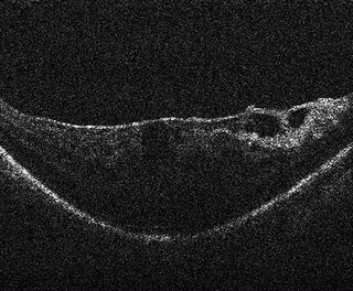
Vitreomacular traction (VMT) and Epiretinal membrane (ERM)
Pre-operative retinal scan shows vitreomacular traction (VMT) and Epiretinal membrane (ERM).
OCT reveals residual membrane
After ERM peeling, OCT reveals residual membrane creating longitudinal traction.
OCT confirms that the membrane creating longitudinal traction has been severed
After further surgical intervention, OCT confirms that the membrane creating longitudinal traction has been severed and no macular hole created. The surgeon decides air exchange is not required, sparing the patient from prone positioning.
Contact DB Surgical for More Information.
Republished from Leica Microsystems.