How an Upgradeable Ophthalmic Microscope can Benefit Surgeon and Hospital

Interview with Dr. Seenu M. Hariprasad, University of Chicago


Proveo 8 ophthalmic microscopeMost ophthalmic surgeons agree that optimal visualization is a key criterion when choosing a surgical microscope. However as more and more digital technologies enter the OR, the microscope is becoming more than a pure optical tool.

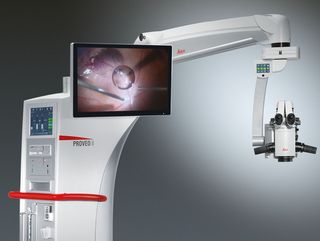
Seenu M. Hariprasad, MD, is the Chief of the Vitreoretinal Service, Director of Clinical Research and Shui-Chin Lee Professor of Ophthalmology and Visual Science at the University of Chicago. He believes the Proveo 8 ophthalmic microscope represents the first of a new generation of ophthalmic microscopes that not only deliver outstanding optical quality, but also serve as a flexible and upgradeable platform for the integration of digital imaging technologies.

What do you see as the advantages of the modular design of the Proveo microscope?

Dr. Hariprasad: Eleven years ago, my hospital purchased a scope for retina surgery that could not be upgraded. In contrast, the Proveo 8 ophthalmic microscope can be used for anterior and posterior surgeries, and expanded and upgraded through time. The ability to expand the scope is essentially the ability to extend its life. A facility makes a major investment in a microscope, and when a new tech­nology comes along in a few years, a new component can be added to the scope. If the scope is not expandable and cannot accept the new compo­nent, then the facility has to replace the scope at a much greater expense.

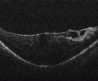
With rapidly evolving medical technologies, we do not know what will come along — even in the near future — so it makes sense to have the ability to expand. One example is intrasurgical OCT, an exciting new devel­opment from a retina perspective that is in its infancy today. The Proveo 8 microscope is built to accept an integrated OCT component for future integration. As well as the possibility to upgrade with future developments, the microscope also enables sophisticated recording devices, Toric Alignment Navigation, and 3D viewing to be added at any time.

The Proveo 8 microscope is designed for anterior and posterior surgeries – what benefit does that offer you and your hospital?

Dr. Hariprasad: The versatility for different ophthal­mic surgeons is an enormous advan­tage because it doubles the potential use and return on a large invest­ment, while potentially eliminating the need for two microscopes, depending on the setting and volume. Hospitals are happy to get something that all doctors can use.

How would you sum up the advantages of the Proveo 8 ophthalmic microscope?

Dr. Hariprasad: As a retina surgeon, I rely on my ophthalmic microscope for high-resolution visualization during surgery. I’m excited about the Proveo product as it offers the latest visualization innovations while also allowing existing and future digital imaging technologies to be integrated into a sleek, modern design. A range of next-generation features may well make the Proveo 8 an essential scope for posterior segment surgeons.


Contact DB Surgical for More Information.
Republished from Leica Microsystems.

How Intrasurgical OCT can Impact the Surgical Approach to Membrane Peeling

OCTWhen treating patients with vitreomacular traction (VMT), peeling of the inner limiting membrane (ILM) or an epiretinal membrane (ERM) is often indicated. Successful outcomes depend on precise surgical maneuvers as well as the surgeon’s experience in order to assess whether the entire membrane has been removed, as this is crucial for successful release of traction. Having as much visual information as possible helps the surgeon confidently perform membrane peeling. It also supports identification of residual membranes, and examination of the retinal morphology immediately following the procedure for complications such as macular holes, sub-retinal edema or residual traction. High-resolution cross-sectional imaging provided by intrasurgical optical coherence tomography (OCT) can provide vital anatomic information to surgeons during retinal procedures, helping to guide surgical decision-making.

Showing different steps of removing an ERM to resolve VMT, the following videos captured with EnFocus intrasurgical Optical Coherence Tomography demonstrate how visualization of the membrane can impact the surgical process of membrane peeling. Imaging with EnFocus intrasurgical OCT supports identification of an epiretinal membrane (ERM) causing vitreomacular traction (VMT) and helps guide intrasurgical decision-making during membrane peeling:


Vitreomacular traction (VMT) and Epiretinal membrane (ERM)

Pre-operative retinal scan shows vitreomacular traction (VMT) and Epiretinal membrane (ERM).

 OCT reveals residual membrane

After ERM peeling, OCT reveals residual membrane creating longitudinal traction.

OCT confirms that the membrane creating longitudinal traction has been severed

After further surgical intervention, OCT confirms that the membrane creating longitudinal traction has been severed and no macular hole created. The surgeon decides air exchange is not required, sparing the patient from prone positioning.


Contact DB Surgical for More Information.

Republished from Leica Microsystems.


The Impact of a Stable Red Reflex and Enhanced Focus in Cataract Surgery


Interview with Ike Ahmed: “I was captivated by Proveo’s unparalleled and consistent red reflex throughout the entire procedure.”

  • Would you please explain your field of work and describe the challenges you are dealing with?
  • What importance does the red reflex for cataract surgery have? In which way does Proveo 8 make a difference?
  • What impact does the quality of the assistant’s view have on the surgery?
  • Do technologies like IOL guidance systems or intraoperative OCT play a role in your OR?
  • If you summarize your experience with Proveo 8 in one sentence, what would you say?

Would you please explain your field of work and describe the challenges you are dealing with?

Ike Ahmed: My focus lies primarily on complicated cataract cases, lens implants and glaucoma surgery. I have an academic practice, I am teaching residence fellows and medical students regularly. I am also very actively involved in research and innovation in medical technology and education as well. In the three different centers within the greater Toronto area where I operate I am working with different microscopes, different technologies and surgical tools.

My interest lies as well in the development of surgical instrumentation and some of my own devices for lens implants as well. I really take a lot of interest and pride in optimizing the surgical field as best possible, which includes the documentation and recording of cases to learn from and to teach.

One of the things in eye surgery which is most challenging is that we have to see what we are doing. In some cases this can be difficult, especially in complicated cataract or glaucoma procedures. Visualization can be challenging because of the view or because of the pathology.

Enhancing technology can help us to achieve optimal visualization, whether that is with microscopes or with one of the newer technologies like 3D heads up display, surgical guidance systems and OCT guided procedures. I am fascinated by the possibility to expand our work beyond what we can see with a basic microscope set up!

What importance does the red reflex for cataract surgery have? In which way does Proveo 8 make a difference?

Ike Ahmed: One of the challenges with visualization is enhancing the red reflex particularly during phacoelmusification, cataract extraction and also during the intraocular lens surgery. The red reflex gives us the ideal contrast as well as the important depth in terms of where we are working within the eye.

So one of the important features of a microscope is to ensure the red reflex and optimal visualization throughout the entire procedure – not only during capsulorhexis, but during lens extraction as well.

One of the benefits especially with Proveo 8 is the way the illumination is achieved by using four coaxial LED light sources. The optics of the microscope and the innovative additional depth of focus allow us to enhance the ability to visualize the procedure throughout the entire case.

That is the experience I had with the Proveo 8. I appreciate the benefit of having the enhanced visualization whether it means of the red reflex or it means of the tissue itself. Optimizing the quality of view is one thing that is beneficial.

But there is another thing that I think is pretty cool: I like to zoom in to see what I am doing with a high magnification. Normally, that means frequent refocusing. But the enhanced focusing features of the Proveo 8 microscope, which is called FusionOptics, allows us to maintain focus over a larger depth – which means we are working with the foot pedal less, we can see more, and we can do better surgery. I found that helps.

What impact does the quality of the assistant’s view have on the surgery?

Ike Ahmed: I always have an assistant whether a resident or a fellow working with me. I am also often assisting them during surgery and so it is absolutely critical that the quality of the assistant’s scope is the same as the surgeon’s scope.

By having maintenance of the red reflex and having magnification linked with the surgeon ensures that I have the optimal teaching environment. That is one thing I really liked and enjoyed with the microscope. It is important with all microscopes in terms of what I can do as far as teaching and visualizing.

Do technologies like IOL guidance systems or intraoperative OCT play a role in your OR?

Ike Ahmed: We are still early with it. The technological merger of diagnostics and microscope – because I think that’s where we are going with this – leads to enhanced workflow and accuracy. But we are going to learn more about where we are going with this.

The OCT for retina and cornea is a nice way to determine tissue plane and optimal dissection. In cataract/IOL surgery we are looking at this for optimal lens positioning. Its use for angle visualization is potentially cool for glaucoma surgery. We are still early in this and I am excited where this may go.

If you summarize your experience with Proveo 8 in one sentence, what would you say?

Ike Ahmed: It was pretty mind blowing – I was captivated by the unparalleled and consistent red reflex and tissue visualization throughout the entire procedure.


Contact DB Surgical for more information.


Cataract Surgery with CoAx4 Illumination

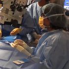
A stable red reflex is one of the most important features of an ophthalmic surgical microscope for cataract surgery. It’s the red reflex that makes the structure of the lens visible and thus makes for an uncompromised view for a successful and secure surgery. One of the challenges with visualization is enhancing the red reflex particularly during phacoelmusification, cataract extraction and also during the intraocular lens surgery.

However, conventional red reflex illumination often decreases during the critical phases of the procedure like phacoemulsification. A new illumination technology with four individual beam paths overcomes these drawbacks.

The Proveo 8 ophthalmic microscope is the first system to feature the new technology. The CoAx4 Illumination provides a stable and consistent red reflex throughout the entire surgical procedure. Both main surgeon and assistant share the same view and full red reflex.

Cataract surgeons rely on a red reflex

The red reflex is the reddish-orange reflection from the retina that is observed through an ophthalmic microscope as coaxial light goes into and out of the pupil of the eye. Cataract surgeons rely on the red reflex and depth of field to precisely view the elements in the eye during each step of surgery, such as corneal incisions, division of nuclear material, removing the cortex, cleaning of the anterior and posterior leaves of the capsular bag, and placing the IOL.

Consistent visualization is especially important when performing capsulorhexis which is best judged by the shadow its edge creates with the red reflex. The brighter and more even the red reflex is, the more distinct the capsular edge becomes. This enhances surgical ease in performing centered and appropriately sized capsulorhexis in all types of cataract.

Consequences of a diminished red reflex

As the eye moves or the position of the microscope is changed, the red reflex may decrease, making certain phases of cataract surgery more difficult, even dangerous. For example, with a diminished red reflex it is more difficult to create a round and appropriately sized capsulorhexis, and to determine where within the cataract the phaco tip is being placed.

Most notably, without consistent illumination it is more difficult to view remaining nuclear material in the capsular bag during phacoemulsificaton and the posterior capsule bag during the procedure.

Four beam paths facilitate stable red reflex

A new technology for an ophthalmic microscope makes it possible to work with a stable and consistent red reflex. Four individual coaxial illumination beam paths down to the patient eye with a perpendicular entrance to the retina are providing a straight red reflex throughout the entire procedure. Compared to known microscopes, the image appears equal for all possible observers, the main observer, the assistant and the documentation.

Stereo red reflex for all observers

The CoAx4 Illumination is the only technology that makes the red reflex fully visible for both main surgeon and assistant. Surgeon and assistant sharing the same view of the surgical field with excellent contrast and red reflex makes cataract surgery more secure and is a valuable asset to teaching.

Adjustable illumination diameter enhances contrast

The Proveo 8 ophthalmic microscope is the first system to feature the new technology. With an adjustable illumination diameter sizeable from 4 – 23 mm the surgeon can choose via footswitch if he needs to illuminate the whole eye or prefers to match the field of illumination to the pupil size. A reduced illumination diameter produces less sclera reflections and offers more contrast, while a wider illumination is more flexible with respect to patient’s movements.

The CoAx4 Illumination also provides a camera image that is identical to the optical view through the ocular. The light for the camera is taken from the assistant optical path and thus leaves the main surgeon’s view with 100% of the light.


Contact DB Surgical for more information.

Republished from Leica Microsystems

Video Talk by Sonia H. Yoo: Interoperative OCT Imaging of Selective Lamellar Keratoplasty

The work prepared and presented within this video demonstrates preliminary research, and the promising future of perioperative OCT to aid the diagnosis of physiological and pathological conditions during ophthalmic surgery in order to help guide surgical decision making in anterior segment cases.

Showing SDOCT Images acquired with an Envisu SD-OCT system, Prof. Yoo demonstrates the use of interoperative OCT imaging in several important surgical procedures such as DALK, DSAEK and DMEK.

Republished from Leica Microsystems