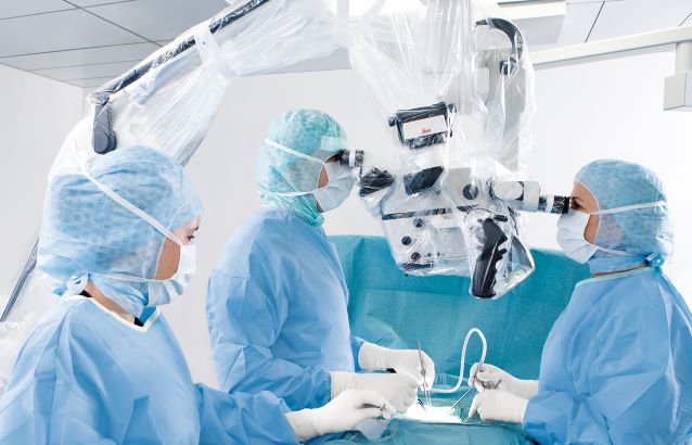
– Adhering to the highest industry and safety standards for operating surgical technology.
All rental microscopes are offered on both a short-term and long-term basis and come complete with installation and case coverage by a certified representative.
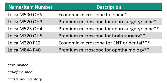
See below for the list of surgical microscopes that DB Surgical currently has available for rent throughout the Southeastern U.S.
To learn more, contact your representative here: Contact Us
To obtain information on pricing and availability, submit a request here: Quote Request
Interview with Dr. Seenu M. Hariprasad, University of Chicago
Most ophthalmic surgeons agree that optimal visualization is a key criterion when choosing a surgical microscope. However as more and more digital technologies enter the OR, the microscope is becoming more than a pure optical tool.
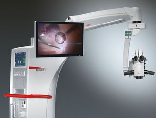
Seenu M. Hariprasad, MD, is the Chief of the Vitreoretinal Service, Director of Clinical Research and Shui-Chin Lee Professor of Ophthalmology and Visual Science at the University of Chicago. He believes the Proveo 8 ophthalmic microscope represents the first of a new generation of ophthalmic microscopes that not only deliver outstanding optical quality, but also serve as a flexible and upgradeable platform for the integration of digital imaging technologies.
What do you see as the advantages of the modular design of the Proveo microscope?
Dr. Hariprasad: Eleven years ago, my hospital purchased a scope for retina surgery that could not be upgraded. In contrast, the Proveo 8 ophthalmic microscope can be used for anterior and posterior surgeries, and expanded and upgraded through time. The ability to expand the scope is essentially the ability to extend its life. A facility makes a major investment in a microscope, and when a new technology comes along in a few years, a new component can be added to the scope. If the scope is not expandable and cannot accept the new component, then the facility has to replace the scope at a much greater expense.
With rapidly evolving medical technologies, we do not know what will come along — even in the near future — so it makes sense to have the ability to expand. One example is intrasurgical OCT, an exciting new development from a retina perspective that is in its infancy today. The Proveo 8 microscope is built to accept an integrated OCT component for future integration. As well as the possibility to upgrade with future developments, the microscope also enables sophisticated recording devices, Toric Alignment Navigation, and 3D viewing to be added at any time.
The Proveo 8 microscope is designed for anterior and posterior surgeries – what benefit does that offer you and your hospital?
Dr. Hariprasad: The versatility for different ophthalmic surgeons is an enormous advantage because it doubles the potential use and return on a large investment, while potentially eliminating the need for two microscopes, depending on the setting and volume. Hospitals are happy to get something that all doctors can use.
How would you sum up the advantages of the Proveo 8 ophthalmic microscope?
Dr. Hariprasad: As a retina surgeon, I rely on my ophthalmic microscope for high-resolution visualization during surgery. I’m excited about the Proveo product as it offers the latest visualization innovations while also allowing existing and future digital imaging technologies to be integrated into a sleek, modern design. A range of next-generation features may well make the Proveo 8 an essential scope for posterior segment surgeons.
To achieve the best possible patient outcomes, surgeons have to remain focused through every step of every surgery. The Leica M530 OH6 supports surgeons to achieve this by providing:
Outstanding visualization and imaging technology
In an upgradeable, ergonomic surgical microscope
Complete clarity for rear assistant
Independent fine focus adjustment for the rear assistant ensures optimal visualization
Smoothly maneuverable
Effortless handling with full range of movement and tilt of the optics carrier for adaptation to different applications
VIDEO: Joshua Bederson, M.D., utilizes the latest simulation and virtual reality advances during neurosurgery.
Credit: Mount Sinai Health System
Joshua Bederson, MD, Professor and System Chair for the Department of Neurosurgery at Mount Sinai Health System, is the first neurosurgeon to use CaptiView – a microscope image injection system from Leica Microsystems that overlays critical virtual reality imaging directly onto the brain when viewed through the eyepiece, known as the ocular, during surgery. This new microscope technology allows images of chosen objects, including original CT, MRI and angiogram datasets, to be superimposed, or ‘injected,’ directly into the neurosurgeon’s eyepiece during microscopic surgery.
“This next-generation augmented virtual reality tool provides real-time information in ways never before realized,” says Dr. Bederson, who is now using the technology for all of his cases. He worked closely with Leica Microsystems and Brainlab® to develop the surgical navigation tool.
The CaptiView image injection system utilizes Brainlab® Cranial 3.1 Navigation Software in conjunction with a Leica M530 OH6 microscope. The heads-up display provides neurovascular and fiber-track information in 2D or 3D as well as on-screen video overlays visible through the ocular. The microscope integration also allows the surgeon to switch views in the eyepiece, toggling between live and pre-operative anatomical images using handle control buttons or footswitch for ease of use and uninterrupted workflow. Markers attached to the microscope enable positional tracking and autofocus.
This new technology will be utilized alongside Surgical Navigation Advanced Platform (SNAP) developed by Surgical Theater, LLC, which is a standard feature in the operating room. SNAP provides advanced 3D visualization technology that gives surgeons an intraoperative and patient-specific 3D environment to plan and understand surgical approaches.
“We are driving and advancing the development of next-generation simulation and virtual reality technology, which can help improve patient outcomes and solve neurosurgical challenges,” says Dr. Bederson.
Dr. Bederson is an expert in skull-base and cerebrovascular surgery and has performed more than 3,600 neurosurgical operations at Mount Sinai. Dr. Bederson owns equity in Surgical Theater, LLC.
Video case study & whitepaper
Costas G. Hadjipanayis, MD, PhD
Mount Sinai Beth Israel, Department of Neurosurgery, New York, NY, USA
Neurosurgical intervention remains the first step in malignant glioma management and is an important prognostic factor in this patient population. Completeness of resection is a significant, independent predictor of survival, but accurate discrimination between tumor and normal brain tissue is challenging.
Although many studies confirm that near-complete resection of contrast-enhancing tumor is necessary to positively affect overall survival, even “complete” resections routinely fail to fully remove the tumor’s infiltration zone. Because of these and other challenges, 96% of all tumors recur within their former resection margin—typically within 7 to 10 months of primary surgery.
With the existing limitations of neuronavigation in complete glioma resection surgery, better intraoperative visualization is needed to minimize obstacles and, ideally, lead to better patient outcomes.
In this application note, Dr. Hadjipanayis explains:
Limitations of neuronavigation
The biochemistry behind Gliolan®
Why Gliolan® (5-ALA) is a better solution
Paradigm shifts that can change the course of treatment
Utility with specific tumor types
Efficacy: Making a difference in patient outcomes
Clinical benefits of Gliolan (5-ALA)
In this video case study, Dr. Hadjipanayis used Gliolan to perform fluorescence guided surgery on a patient with a malignant brain tumor. The surgery was performed using a Leica M530 OH6 neurosurgical microscope with the FL400* intraoperative fluorescence system:
* For all fluorescence modules, please check the status of regulatory approval for your country with your local Leica Microsystems representative.
In cataract surgery, ophthalmologists rely on the red reflex which provides ideal contrast to visualize the capsule, lens and anterior chamber structure. A new illumination technology in the latest of ophthalmic microscopes now appears to provide great breadth of red reflex enhancement throughout the entire procedure.
Dr. Ike Ahmed is one of the first ophthalmologists having tested the Proveo 8 microscope which features the CoAx4 illumination. He is assistant professor and the director of the Glaucoma and Advanced Anterior Segment Surgery fellowship at the University of Toronto, Canada.
Contents
Would you please explain your field of work and describe the challenges you are dealing with?
What importance does the red reflex for cataract surgery have? In which way does Proveo 8 make a difference?
What impact does the quality of the assistant’s view have on the surgery?
Do technologies like IOL guidance systems or intraoperative OCT play a role in your OR?
If you summarize your experience with Proveo 8 in one sentence, what would you say?
Would you please explain your field of work and describe the challenges you are dealing with?
Ike Ahmed: My focus lies primarily on complicated cataract cases, lens implants and glaucoma surgery. I have an academic practice, I am teaching residence fellows and medical students regularly. I am also very actively involved in research and innovation in medical technology and education as well. In the three different centers within the greater Toronto area where I operate I am working with different microscopes, different technologies and surgical tools.
My interest lies as well in the development of surgical instrumentation and some of my own devices for lens implants as well. I really take a lot of interest and pride in optimizing the surgical field as best possible, which includes the documentation and recording of cases to learn from and to teach.
One of the things in eye surgery which is most challenging is that we have to see what we are doing. In some cases this can be difficult, especially in complicated cataract or glaucoma procedures. Visualization can be challenging because of the view or because of the pathology.
Enhancing technology can help us to achieve optimal visualization, whether that is with microscopes or with one of the newer technologies like 3D heads up display, surgical guidance systems and OCT guided procedures. I am fascinated by the possibility to expand our work beyond what we can see with a basic microscope set up!
What importance does the red reflex for cataract surgery have? In which way does Proveo 8 make a difference?
Ike Ahmed: One of the challenges with visualization is enhancing the red reflex particularly during phacoelmusification, cataract extraction and also during the intraocular lens surgery. The red reflex gives us the ideal contrast as well as the important depth in terms of where we are working within the eye.
So one of the important features of a microscope is to ensure the red reflex and optimal visualization throughout the entire procedure – not only during capsulorhexis, but during lens extraction as well.
One of the benefits especially with Proveo 8 is the way the illumination is achieved by using four coaxial LED light sources. The optics of the microscope and the innovative additional depth of focus allow us to enhance the ability to visualize the procedure throughout the entire case.
That is the experience I had with the Proveo 8. I appreciate the benefit of having the enhanced visualization whether it means of the red reflex or it means of the tissue itself. Optimizing the quality of view is one thing that is beneficial.
But there is another thing that I think is pretty cool: I like to zoom in to see what I am doing with a high magnification. Normally, that means frequent refocusing. But the enhanced focusing features of the Proveo 8 microscope, which is called FusionOptics, allows us to maintain focus over a larger depth – which means we are working with the foot pedal less, we can see more, and we can do better surgery. I found that helps.
What impact does the quality of the assistant’s view have on the surgery?
Ike Ahmed: I always have an assistant whether a resident or a fellow working with me. I am also often assisting them during surgery and so it is absolutely critical that the quality of the assistant’s scope is the same as the surgeon’s scope.
By having maintenance of the red reflex and having magnification linked with the surgeon ensures that I have the optimal teaching environment. That is one thing I really liked and enjoyed with the microscope. It is important with all microscopes in terms of what I can do as far as teaching and visualizing.
Do technologies like IOL guidance systems or intraoperative OCT play a role in your OR?
Ike Ahmed: We are still early with it. The technological merger of diagnostics and microscope – because I think that’s where we are going with this – leads to enhanced workflow and accuracy. But we are going to learn more about where we are going with this.
The OCT for retina and cornea is a nice way to determine tissue plane and optimal dissection. In cataract/IOL surgery we are looking at this for optimal lens positioning. Its use for angle visualization is potentially cool for glaucoma surgery. We are still early in this and I am excited where this may go.
If you summarize your experience with Proveo 8 in one sentence, what would you say?
Ike Ahmed: It was pretty mind blowing – I was captivated by the unparalleled and consistent red reflex and tissue visualization throughout the entire procedure.
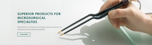
Kirwan Surgical Products, renowned for its pioneering efforts in microsurgery, offers an extensive range of bipolar forceps, each designed to enhance surgical precision and patient outcomes. The key benefits of Kirwan’s bipolar forceps are rooted in their meticulous design and technological sophistication: 1. **Advanced Non-Stick Technology**: Kirwan’s AURA™ and POLARIS line of bipolar forceps incorporates […]