Interview with Dr. Seenu M. Hariprasad, University of Chicago
 Most ophthalmic surgeons agree that optimal visualization is a key criterion when choosing a surgical microscope. However as more and more digital technologies enter the OR, the microscope is becoming more than a pure optical tool.
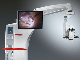
Seenu M. Hariprasad, MD, is the Chief of the Vitreoretinal Service, Director of Clinical Research and Shui-Chin Lee Professor of Ophthalmology and Visual Science at the University of Chicago. He believes the Proveo 8 ophthalmic microscope represents the first of a new generation of ophthalmic microscopes that not only deliver outstanding optical quality, but also serve as a flexible and upgradeable platform for the integration of digital imaging technologies.
What do you see as the advantages of the modular design of the Proveo microscope?
Dr. Hariprasad: Eleven years ago, my hospital purchased a scope for retina surgery that could not be upgraded. In contrast, the Proveo 8 ophthalmic microscope can be used for anterior and posterior surgeries, and expanded and upgraded through time. The ability to expand the scope is essentially the ability to extend its life. A facility makes a major investment in a microscope, and when a new technology comes along in a few years, a new component can be added to the scope. If the scope is not expandable and cannot accept the new component, then the facility has to replace the scope at a much greater expense.
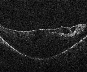
With rapidly evolving medical technologies, we do not know what will come along — even in the near future — so it makes sense to have the ability to expand. One example is intrasurgical OCT, an exciting new development from a retina perspective that is in its infancy today. The Proveo 8 microscope is built to accept an integrated OCT component for future integration. As well as the possibility to upgrade with future developments, the microscope also enables sophisticated recording devices, Toric Alignment Navigation, and 3D viewing to be added at any time.
The Proveo 8 microscope is designed for anterior and posterior surgeries – what benefit does that offer you and your hospital?
Dr. Hariprasad: The versatility for different ophthalmic surgeons is an enormous advantage because it doubles the potential use and return on a large investment, while potentially eliminating the need for two microscopes, depending on the setting and volume. Hospitals are happy to get something that all doctors can use.
How would you sum up the advantages of the Proveo 8 ophthalmic microscope?
Dr. Hariprasad: As a retina surgeon, I rely on my ophthalmic microscope for high-resolution visualization during surgery. I’m excited about the Proveo product as it offers the latest visualization innovations while also allowing existing and future digital imaging technologies to be integrated into a sleek, modern design. A range of next-generation features may well make the Proveo 8 an essential scope for posterior segment surgeons.


