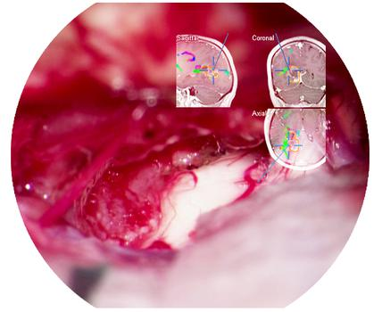
Recording of a brain aneurysm clipping by Vik Udani, MD, Division of Neurosurgery, Senta Clinic, San Diego, CA.
Patient is a 38-year-old female with a history of hypertension and migraines. She was found to have an 8mm unruptured right middle cerebral artery aneurysm and underwent a right frontal temporal craniotomomy for aneurysm clipping.
Dr. Udani on the M530 OH6 surgical microscope: “One of my favorite aspects of the OH6 is the amount of illumination on the surgical field. The light transmission provides sharp, crisp, bright images of fine vascular structures, which greatly enhances my visualization throughout the entire procedure. The improved illumination as well as the integrated camera also provides greater clarity when using the ICG activated filter giving me greater confidence in my surgical decisions.”
Watch this video and experience for yourself: Bright 400-Watt Xenon light system for concentrated light beam into deep cavities; high resolution and depth of focus with exclusive FusionOptics technology.
Image Guided Surgery with microscope image injection aids visualization and orientation during neurosurgery
One of the challenges of neurosurgery is orientation at the surgical site. When resecting tumors, removing arteriovenous malformations, or clipping aneurysms, surgeons often have to work near healthy and functional brain tissue. When resecting the tumor, the challenge is always to spare as much healthy tissue as possible. Neuronavigation technology, also referred to as Image Guided Surgery (IGS) enables surgeons to plan the ideal approach before making a cut and helps to execute that plan by providing intraoperative orientation.
At the base of the human skull there is a particularly high concentration of vital structures: blood vessels, nerves, centers for hormonal control of bodily functions, and centers for regulating breathing, blood pressure, and heart activity. Brain tissue cannot regenerate itself, so even the smallest injury can cause irreversible, profound brain damage, leaving patients with a permanent disability. This also makes the treatment of disease very difficult. Malignant gliomas, which account for 45% to 50% of all brain tumors, infiltrate into brain tissue as they grow. Even under a surgical microscope, peripheral tumor regions are hard to differentiate from healthy tissue.
Continuous real-time view of the surgical field
Neuronavigation works like a GPS system, enabling accurate position tracking of instruments within the patient’s anatomy. Surgeons can outline the tumor, identify organs at risk, and define the optimal access pathway to the tumor to prevent injury to healthy structures.
Before surgery, CT or MR images of the patient are taken. In the operating room, the navigation system matches these images up with the live patient images using a wireless laser pointer. The pointer identifies surface points on the patient’s skin which are transmitted to the navigation system via an infrared camera. During the procedure, the camera also transmits the locations of the instruments and equipment – including the microscope – which are outfitted with reflective markers.
The patient images prepared prior to surgery are loaded into the navigation system and reconstructed into images on the monitor or injected into the oculars of the surgical microscope. This gives the surgeon a continuous, real-time map of the position of the instruments in relation to the brain structures and the tumor.
Image injection for a complete picture
To view the neuronavigation data, surgeons normally need to raise their heads from the microscope to look at the navigation screen. Thanks to image injection systems, like the new CaptiView from Leica, and in combination with the new picture-in-picture feature from Brainlab, the surgeon can see navigation data directly in the microscope’s eyepieces. The full-HD display of CaptiView image injection with LED backlighting provides bright, crisp injected images with distinct margins. Neuronavigation data from the Image Guided Surgery system can be viewed either in the right, the left, or both eyes and the CaptiView system is also able to display real-time HD images from an endoscope and FL800 vascular fluorescence. ”By having all information directly in the microscope’s ocular, the surgeon does not have to reorient between the microscope and the navigation screen, but can remain fully concentrated on the patient during the whole procedure”, explains David A Smith, Product Manager at Leica Microsystems. Furthermore, injected images are also displayed in the assistant binoculars and can be recorded in full HD quality, supporting teaching both inside and outside of the OR.
Microscope integration offers new imaging options
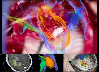
Brainlab is one of the market leaders in the field of image-guided technology and was the first to work together with Leica to integrate its neuronavigation software with surgical microscopes. The latest microscope integration software from Brainlab works seamlessly with CaptiView image injection and M530 microscopes from Leica allowing data to be injected into the microscope’s eyepiece and additionally displayed on screen. With improved 3D visualization, the new Brainlab software provides a more realistic representation of planned objects, structure and fiber tracts throughout the entire procedure.
Touch-based rotation of the microscope view on the navigation screen allows for a look behind the actual video plane to reveal underlying structures in 3D for more anatomical insight. The “Probe’s Eye” feature reconstructs the patient dataset to the microscope’s position and orientation for a direct comparison of preoperative data and intraoperative anatomy. With “Navigation update” the surgeon is able to correct initial registration based on the patient’s anatomical landmarks.
Limiting guesswork in tumor resection
Every neurosurgical procedure demands a vast amount of surgeon experience, patience and competence. Although neuronavigation does not replace surgical skill, it effectively supports the surgeon in pre-operative planning and during the procedure by giving more precise information in the most critical moments.
“The integration of the microscope with neuronavigation helps the surgeon to accurately identify questionable areas”, explains Valentin Elefteriu, Product Manager Augmented Reality at Brainlab. “For example, if they focus on the edge of a tumor and can’t tell exactly where the tumor tissue ends, a look at the pre-planned anatomic data with microscope image overlay reveals whether the focused area is part of the tumor or not. Precise planning may also minimize head shaving, skin incisions, and bone flap sawing and thus reduce stress for the patient.”
Brainlab is a registered trademark of Brainlab AG registered in Europe, the United States and other countries.
VIDEO: Joshua Bederson, M.D., utilizes the latest simulation and virtual reality advances during neurosurgery.
Credit: Mount Sinai Health System
Joshua Bederson, MD, Professor and System Chair for the Department of Neurosurgery at Mount Sinai Health System, is the first neurosurgeon to use CaptiView – a microscope image injection system from Leica Microsystems that overlays critical virtual reality imaging directly onto the brain when viewed through the eyepiece, known as the ocular, during surgery. This new microscope technology allows images of chosen objects, including original CT, MRI and angiogram datasets, to be superimposed, or ‘injected,’ directly into the neurosurgeon’s eyepiece during microscopic surgery.
“This next-generation augmented virtual reality tool provides real-time information in ways never before realized,” says Dr. Bederson, who is now using the technology for all of his cases. He worked closely with Leica Microsystems and Brainlab® to develop the surgical navigation tool.
The CaptiView image injection system utilizes Brainlab® Cranial 3.1 Navigation Software in conjunction with a Leica M530 OH6 microscope. The heads-up display provides neurovascular and fiber-track information in 2D or 3D as well as on-screen video overlays visible through the ocular. The microscope integration also allows the surgeon to switch views in the eyepiece, toggling between live and pre-operative anatomical images using handle control buttons or footswitch for ease of use and uninterrupted workflow. Markers attached to the microscope enable positional tracking and autofocus.
This new technology will be utilized alongside Surgical Navigation Advanced Platform (SNAP) developed by Surgical Theater, LLC, which is a standard feature in the operating room. SNAP provides advanced 3D visualization technology that gives surgeons an intraoperative and patient-specific 3D environment to plan and understand surgical approaches.
“We are driving and advancing the development of next-generation simulation and virtual reality technology, which can help improve patient outcomes and solve neurosurgical challenges,” says Dr. Bederson.
Dr. Bederson is an expert in skull-base and cerebrovascular surgery and has performed more than 3,600 neurosurgical operations at Mount Sinai. Dr. Bederson owns equity in Surgical Theater, LLC.
Kirwan Surgical Products, renowned for its pioneering efforts in microsurgery, offers an extensive range of bipolar forceps, each designed to enhance surgical precision and patient outcomes. The key benefits of Kirwan’s bipolar forceps are rooted in their meticulous design and technological sophistication: 1. **Advanced Non-Stick Technology**: Kirwan’s AURA™ and POLARIS line of bipolar forceps incorporates […]

 One of the challenges of neurosurgery is orientation at the surgical site. When resecting tumors, removing arteriovenous malformations, or clipping aneurysms, surgeons often have to work near healthy and functional brain tissue. When resecting the tumor, the challenge is always to spare as much healthy tissue as possible. Neuronavigation technology, also referred to as Image Guided Surgery (IGS) enables surgeons to plan the ideal approach before making a cut and helps to execute that plan by providing intraoperative orientation.