Discover the Leica M530 OH6 Surgical Microscope with FusionOptics

Leica M530 OH6 Neurosurgery Microscope

To achieve the best possible patient outcomes, surgeons have to remain focused through every step of every surgery.  The Leica M530 OH6 supports surgeons to achieve this by providing:

  •  Outstanding visualization and imaging technology
    • In an upgradeable, ergonomic surgical microscope
  • Complete clarity for rear assistant
    • Independent fine focus adjustment for the rear assistant ensures optimal visualization
  • Smoothly maneuverable
    • Effortless handling with full range of movement and tilt of the optics carrier for adaptation to different applications

Contact DB Surgical for more information.

Republished from Leica Microsystems.

Microscope Imaging System Integrates Virtual Reality Technology

VIDEO: Joshua Bederson, M.D., utilizes the latest simulation and virtual reality advances during neurosurgery.
Credit: Mount Sinai Health System


Joshua Bederson, MD, Professor and System Chair for the Department of Neurosurgery at Mount Sinai Health System, is the first neurosurgeon to use CaptiView – a microscope image injection system from Leica Microsystems that overlays critical virtual reality imaging directly onto the brain when viewed through the eyepiece, known as the ocular, during surgery. This new microscope technology allows images of chosen objects, including original CT, MRI and angiogram datasets, to be superimposed, or ‘injected,’ directly into the neurosurgeon’s eyepiece during microscopic surgery.

“This next-generation augmented virtual reality tool provides real-time information in ways never before realized,” says Dr. Bederson, who is now using the technology for all of his cases. He worked closely with Leica Microsystems and Brainlab® to develop the surgical navigation tool.

The CaptiView image injection system utilizes Brainlab® Cranial 3.1 Navigation Software in conjunction with a Leica M530 OH6 microscope. The heads-up display provides neurovascular and fiber-track information in 2D or 3D as well as on-screen video overlays visible through the ocular. The microscope integration also allows the surgeon to switch views in the eyepiece, toggling between live and pre-operative anatomical images using handle control buttons or footswitch for ease of use and uninterrupted workflow. Markers attached to the microscope enable positional tracking and autofocus.

This new technology will be utilized alongside Surgical Navigation Advanced Platform (SNAP) developed by Surgical Theater, LLC, which is a standard feature in the operating room. SNAP provides advanced 3D visualization technology that gives surgeons an intraoperative and patient-specific 3D environment to plan and understand surgical approaches.

“We are driving and advancing the development of next-generation simulation and virtual reality technology, which can help improve patient outcomes and solve neurosurgical challenges,” says Dr. Bederson.

Dr. Bederson is an expert in skull-base and cerebrovascular surgery and has performed more than 3,600 neurosurgical operations at Mount Sinai. Dr. Bederson owns equity in Surgical Theater, LLC.


Contact DB Surgical for more information.

Republished from EurekAlert!

If it Glows, it’s Tumor – 5-ALA Fluorescence for More Accurate Intraoperative Visualization

Video case study & whitepaper
Costas G. Hadjipanayis, MD, PhD
Mount Sinai Beth Israel, Department of Neurosurgery, New York, NY, USA


p1Neurosurgical intervention remains the first step in malignant glioma management and is an important prognostic factor in this patient population. Completeness of resection is a significant, independent predictor of survival, but accurate discrimination between tumor and normal brain tissue is challenging.

Although many studies confirm that near-complete resection of contrast-enhancing tumor is necessary to positively affect overall survival, even “complete” resections routinely fail to fully remove the tumor’s infiltration zone. Because of these and other challenges, 96% of all tumors recur within their former resection margin—typically within 7 to 10 months of primary surgery.

With the existing limitations of neuronavigation in complete glioma resection surgery, better intraoperative visualization is needed to minimize obstacles and, ideally, lead to better patient outcomes.

In this application note, Dr. Hadjipanayis explains:

  • Limitations of neuronavigation
  • The biochemistry behind Gliolan®
  • Why Gliolan® (5-ALA) is a better solution
  • Paradigm shifts that can change the course of treatment
  • Utility with specific tumor types
  • Efficacy: Making a difference in patient outcomes
  • Clinical benefits of Gliolan (5-ALA)

In this video case study, Dr. Hadjipanayis used Gliolan to perform fluorescence guided surgery on a patient with a malignant brain tumor. The surgery was performed using a Leica M530 OH6 neurosurgical microscope with the FL400* intraoperative fluorescence system:

* For all fluorescence modules, please check the status of regulatory approval for your country with your local Leica Microsystems representative.


Contact DB Surgical for more information.

Republished from Leica Microsystems.


The Impact of a Stable Red Reflex and Enhanced Focus in Cataract Surgery


Interview with Ike Ahmed: “I was captivated by Proveo’s unparalleled and consistent red reflex throughout the entire procedure.”

  • Would you please explain your field of work and describe the challenges you are dealing with?
  • What importance does the red reflex for cataract surgery have? In which way does Proveo 8 make a difference?
  • What impact does the quality of the assistant’s view have on the surgery?
  • Do technologies like IOL guidance systems or intraoperative OCT play a role in your OR?
  • If you summarize your experience with Proveo 8 in one sentence, what would you say?

Would you please explain your field of work and describe the challenges you are dealing with?

Ike Ahmed: My focus lies primarily on complicated cataract cases, lens implants and glaucoma surgery. I have an academic practice, I am teaching residence fellows and medical students regularly. I am also very actively involved in research and innovation in medical technology and education as well. In the three different centers within the greater Toronto area where I operate I am working with different microscopes, different technologies and surgical tools.

My interest lies as well in the development of surgical instrumentation and some of my own devices for lens implants as well. I really take a lot of interest and pride in optimizing the surgical field as best possible, which includes the documentation and recording of cases to learn from and to teach.

One of the things in eye surgery which is most challenging is that we have to see what we are doing. In some cases this can be difficult, especially in complicated cataract or glaucoma procedures. Visualization can be challenging because of the view or because of the pathology.

Enhancing technology can help us to achieve optimal visualization, whether that is with microscopes or with one of the newer technologies like 3D heads up display, surgical guidance systems and OCT guided procedures. I am fascinated by the possibility to expand our work beyond what we can see with a basic microscope set up!

What importance does the red reflex for cataract surgery have? In which way does Proveo 8 make a difference?

Ike Ahmed: One of the challenges with visualization is enhancing the red reflex particularly during phacoelmusification, cataract extraction and also during the intraocular lens surgery. The red reflex gives us the ideal contrast as well as the important depth in terms of where we are working within the eye.

So one of the important features of a microscope is to ensure the red reflex and optimal visualization throughout the entire procedure – not only during capsulorhexis, but during lens extraction as well.

One of the benefits especially with Proveo 8 is the way the illumination is achieved by using four coaxial LED light sources. The optics of the microscope and the innovative additional depth of focus allow us to enhance the ability to visualize the procedure throughout the entire case.

That is the experience I had with the Proveo 8. I appreciate the benefit of having the enhanced visualization whether it means of the red reflex or it means of the tissue itself. Optimizing the quality of view is one thing that is beneficial.

But there is another thing that I think is pretty cool: I like to zoom in to see what I am doing with a high magnification. Normally, that means frequent refocusing. But the enhanced focusing features of the Proveo 8 microscope, which is called FusionOptics, allows us to maintain focus over a larger depth – which means we are working with the foot pedal less, we can see more, and we can do better surgery. I found that helps.

What impact does the quality of the assistant’s view have on the surgery?

Ike Ahmed: I always have an assistant whether a resident or a fellow working with me. I am also often assisting them during surgery and so it is absolutely critical that the quality of the assistant’s scope is the same as the surgeon’s scope.

By having maintenance of the red reflex and having magnification linked with the surgeon ensures that I have the optimal teaching environment. That is one thing I really liked and enjoyed with the microscope. It is important with all microscopes in terms of what I can do as far as teaching and visualizing.

Do technologies like IOL guidance systems or intraoperative OCT play a role in your OR?

Ike Ahmed: We are still early with it. The technological merger of diagnostics and microscope – because I think that’s where we are going with this – leads to enhanced workflow and accuracy. But we are going to learn more about where we are going with this.

The OCT for retina and cornea is a nice way to determine tissue plane and optimal dissection. In cataract/IOL surgery we are looking at this for optimal lens positioning. Its use for angle visualization is potentially cool for glaucoma surgery. We are still early in this and I am excited where this may go.

If you summarize your experience with Proveo 8 in one sentence, what would you say?

Ike Ahmed: It was pretty mind blowing – I was captivated by the unparalleled and consistent red reflex and tissue visualization throughout the entire procedure.


Contact DB Surgical for more information.


Cataract Surgery with CoAx4 Illumination

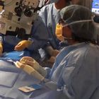
A stable red reflex is one of the most important features of an ophthalmic surgical microscope for cataract surgery. It’s the red reflex that makes the structure of the lens visible and thus makes for an uncompromised view for a successful and secure surgery. One of the challenges with visualization is enhancing the red reflex particularly during phacoelmusification, cataract extraction and also during the intraocular lens surgery.

However, conventional red reflex illumination often decreases during the critical phases of the procedure like phacoemulsification. A new illumination technology with four individual beam paths overcomes these drawbacks.

The Proveo 8 ophthalmic microscope is the first system to feature the new technology. The CoAx4 Illumination provides a stable and consistent red reflex throughout the entire surgical procedure. Both main surgeon and assistant share the same view and full red reflex.

Cataract surgeons rely on a red reflex

The red reflex is the reddish-orange reflection from the retina that is observed through an ophthalmic microscope as coaxial light goes into and out of the pupil of the eye. Cataract surgeons rely on the red reflex and depth of field to precisely view the elements in the eye during each step of surgery, such as corneal incisions, division of nuclear material, removing the cortex, cleaning of the anterior and posterior leaves of the capsular bag, and placing the IOL.

Consistent visualization is especially important when performing capsulorhexis which is best judged by the shadow its edge creates with the red reflex. The brighter and more even the red reflex is, the more distinct the capsular edge becomes. This enhances surgical ease in performing centered and appropriately sized capsulorhexis in all types of cataract.

Consequences of a diminished red reflex

As the eye moves or the position of the microscope is changed, the red reflex may decrease, making certain phases of cataract surgery more difficult, even dangerous. For example, with a diminished red reflex it is more difficult to create a round and appropriately sized capsulorhexis, and to determine where within the cataract the phaco tip is being placed.

Most notably, without consistent illumination it is more difficult to view remaining nuclear material in the capsular bag during phacoemulsificaton and the posterior capsule bag during the procedure.

Four beam paths facilitate stable red reflex

A new technology for an ophthalmic microscope makes it possible to work with a stable and consistent red reflex. Four individual coaxial illumination beam paths down to the patient eye with a perpendicular entrance to the retina are providing a straight red reflex throughout the entire procedure. Compared to known microscopes, the image appears equal for all possible observers, the main observer, the assistant and the documentation.

Stereo red reflex for all observers

The CoAx4 Illumination is the only technology that makes the red reflex fully visible for both main surgeon and assistant. Surgeon and assistant sharing the same view of the surgical field with excellent contrast and red reflex makes cataract surgery more secure and is a valuable asset to teaching.

Adjustable illumination diameter enhances contrast

The Proveo 8 ophthalmic microscope is the first system to feature the new technology. With an adjustable illumination diameter sizeable from 4 – 23 mm the surgeon can choose via footswitch if he needs to illuminate the whole eye or prefers to match the field of illumination to the pupil size. A reduced illumination diameter produces less sclera reflections and offers more contrast, while a wider illumination is more flexible with respect to patient’s movements.

The CoAx4 Illumination also provides a camera image that is identical to the optical view through the ocular. The light for the camera is taken from the assistant optical path and thus leaves the main surgeon’s view with 100% of the light.


Contact DB Surgical for more information.

Republished from Leica Microsystems

You Can’t Treat What You Can’t See: Ophthalmic Microscope Proveo 8

In the most critical moments of ophthalmic surgery, you need to be able to rely on consistent, uncompromised images, because you can’t treat what you can’t see.

The Proveo 8 ophthalmic microscope goes beyond conventional visualization. Its exclusive optical technology provides you with both constant red reflex and a rich texture view, throughout entire anterior and posterior procedures.

Proveo 8 pushes the boundaries of visualization:

  • CoAx4 Illumination technology for enhanced view during all cataract surgery stages, including phacoemulsification
  • FusionOptics technology for better texture view in retina and cataract surgery

Contact DB Surgical for more information.

Republished from Leica Microsystems

Leica Microsystems Receives Award as Technology Leader

Wetzlar, Germany. The research and consulting company Frost & Sullivan has recognized Leica Microsystems’ newly acquired Optical Coherence Tomography (OCT) company Bioptigen of Morrisville, North Carolina, USA, as a technology leader and pioneer of the North American ophthalmic imaging industry. The Frost & Sullivan Award ceremony took place January 13, 2016 in San Diego, USA.

In their appraisal, Frost & Sullivan calls attention to the value represented in Bioptigen’s OCT solutions: “Frost & Sullivan recognizes the enormous value addition Bioptigen’s Envisu Spectral Domain (SD)-OCT platform brings to ophthalmic imaging.”

Particularly emphasized are the highly customizable imaging solutions for pre-clinical and clinical applications. Bioptigen is offering hand-held OCT imaging capabilities bringing OCT directly to the patient. Quick and sensitive real-time OCT imaging minimizes the challenges of comprehensive eye examinations with supine and non-compliant patients, such as infants and children. Bioptigen offers the world’s only hand-held OCT solution approved by the FDA for use as an aid in the diagnosis of physiological and pathologic conditions of the eye.

Markus Lusser, President of Leica Microsystems, expressed his pride at the award: “Leica Microsystems is honored that Bioptigen has been selected for the Frost & Sullivan Award. It is tangible proof of the Bioptigen team’s dedication to creativity, innovation, and the constant exploration of growth potential in the vision healthcare market.”

Bioptigen develops and manufactures innovative OCT systems and software and provides products for clinical as well as translational research applications globally. Bioptigen currently markets the Envisu OCT for pediatric ophthalmic imaging applications, and a family of OCT imaging products for vision science research. Notably, in December 2015, the company received FDA clearance for EnFocus, an innovative OCT ophthalmic imaging solution to upgrade surgical microscopes with intrasurgical OCT capabilities directly integrated in the light path of the microscope. Bioptigen was acquired by Leica Microsystems in 2015.


Contact DB Surgical for more information.

Republished from Leica Microsystems


Leica Microsystems’ Subsidiary Bioptigen Receives FDA’s 510(k) Clearance for EnFocus

Morrisville, NC, USA. Leica Microsystems’ optical coherence tomography (OCT) division Bioptigen received 510(k) clearance from the U.S. Food and Drug Administration (FDA) to market its EnFocus intrasurgical OCT system. EnFocus is an upgrade solution for new and existing ophthalmic surgical microscopes, extending their capability. EnFocus allows visualization of ocular tissue microstructure during ophthalmic surgery in high-resolution, thereby providing both anterior and posterior surgeons with subsurface insight while operating.

“The EnFocus intrasurgical OCT system provides excellent optical imaging performance for the surgeon employing a modular design which enables sharing between multiple microscopes in an operating theater,” said Markus Lusser, President of Leica Microsystems. “It cleverly adds an important capability to ophthalmic microscope platforms. The device is an innovative advancement in ophthalmic imaging that Leica customers called upon us to deliver, similar to the newly introduced Proveo ophthalmic microscope platform.”

“We are very excited to receive clearance to introduce the EnFocus intrasurgical OCT system, which provides ophthalmic surgeons with highly resolved, real-time, deep and wide-field images for use during ophthalmic surgery”, added Eric Buckland, CEO of Bioptigen. “This product introduction builds upon Bioptigen’s 10-year history of delivering advanced OCT imaging tools to the ophthalmic healthcare community”.

The EnFocus imaging systems, while mounted to surgical microscopes, are intended for use with patient populations from pediatric to adults. The systems acquire, process, and display depth-resolved images of ocular tissue using Spectral Domain Optical Coherence Tomography (SD-OCT), and are compatible with common retina fundus viewing systems.


Republished from Leica Microsystems

Leica Microsystems Launches New Ophthalmic Microscope Platform at AAO in Las Vegas

Proveo8-Surgical-Microscope_05

LAS VEGAS and WETZLAR, Germany. Leica Microsystems launches the Proveo 8, the core of a new ophthalmic microscope platform for cataract and vitreoretinal surgeries at the American Academy of Ophthalmology (AAO) Annual Meeting 2015 in Las Vegas, USA.

“‘You can’t treat what you can’t see’, is how a key opinion leader described one of the core challenges facing ophthalmic surgeons today,” says Markus Lusser, President of Leica Microsystems. “This is why we’ve designed our new Proveo platform to deliver exactly what an ophthalmic surgeon needs in order to perform a broad spectrum of ophthalmic procedures with confidence and efficiency. The Proveo 8 accomplishes this by providing consistent, uncompromised red reflex and a crisp, texture-rich image, especially during the most critical moments of each procedure.”

To deliver these advantages, the Proveo 8 is equipped with breakthrough innovations such as FusionOptics and CoAx4 Illumination. FusionOptics is a Leica exclusive technology that combines 40% increased depth of field with high resolution, providing surgeons with a texture-rich image from the edges of a detached membrane to the periphery of the retina, without refocusing. Another Leica exclusive feature, CoAx4 Illumination, delivers light through four individual beam paths and allows for adjustment of the illumination diameter. This results in constant red reflex and high contrast visualization without compromise for main surgeon or the assistant.

“The red reflex of the Proveo 8 is world class, with vividly contrasting edge of the capsulorhexis, but where Proveo has the real edge is in the Phacoemulsification process. Throughout Phaco, the microscope provides optical visibility with contrast, color rendition and crispness better than any other microscope,” says Dr. Ike Ahmed, Head of Ophthalmology at Trillium Health Partners, Mississauga, Ontario, Canada. He also adds: “Proveo is effective in teaching. Its assistant scope presents a full stereo view with excellent contrast and red reflex. Instructors and young surgeons sharing the same field of view accelerates the teaching process.”

The Proveo has been designed to easily integrate innovative surgical technologies like IOL guidance systems for precise IOL positioning, intraoperative OCT to observe ocular tissue microstructures, or 3D “heads-up” surgery. The integrated, yet modular design also allows easy upgrade for additional flexibility. The Proveo therefore provides ophthalmologists with a single platform solution to meet their anterior and posterior imaging needs today and tomorrow.

The American Academy of Ophthalmology (AAO) Annual Meeting 2015 in Las Vegas, USA will take place November 14 to 17, 2015. “We are really excited to unveil our new Proveo platform at AAO,” Lusser says. “With Proveo we developed a future-proof platform that was based on the input of many clinicians worldwide and we can’t wait to deliver the result into the hands of many new customers and contribute to their goal of improving clinical care in the growing ophthalmic surgery market.”

For more information about the Proveo 8 please check http://www.leica-microsystems.com/products/surgical-microscopes/ophthalmology/details/product/leica-proveo-8/

About Leica Microsystems

Leica Microsystems develops and manufactures microscopes and scientific instruments for the analysis of microstructures and nanostructures. Ever since the company started as a family business in the nineteenth century, its instruments have been widely recognized for their optical precision and innovative technology. It is one of the market leaders in compound and stereo microscopy, digital microscopy, confocal laser scanning microscopy with related imaging systems, electron microscopy sample preparation, and surgical microscopes.

Leica Microsystems has seven major plants and product development sites around the world. The company is represented in over 100 countries, has sales and service organizations in 20 countries, and an international network of distribution partners. Its headquarters are located in Wetzlar, Germany.

SOURCE Leica Microsystems