
Video case study & whitepaper
Costas G. Hadjipanayis, MD, PhD
Mount Sinai Beth Israel, Department of Neurosurgery, New York, NY, USA
 Neurosurgical intervention remains the first step in malignant glioma management and is an important prognostic factor in this patient population. Completeness of resection is a significant, independent predictor of survival, but accurate discrimination between tumor and normal brain tissue is challenging.
Although many studies confirm that near-complete resection of contrast-enhancing tumor is necessary to positively affect overall survival, even “complete” resections routinely fail to fully remove the tumor’s infiltration zone. Because of these and other challenges, 96% of all tumors recur within their former resection margin—typically within 7 to 10 months of primary surgery.
With the existing limitations of neuronavigation in complete glioma resection surgery, better intraoperative visualization is needed to minimize obstacles and, ideally, lead to better patient outcomes.
In this application note, Dr. Hadjipanayis explains:
- Limitations of neuronavigation
- The biochemistry behind Gliolan®
- Why Gliolan® (5-ALA) is a better solution
- Paradigm shifts that can change the course of treatment
- Utility with specific tumor types
- Efficacy: Making a difference in patient outcomes
- Clinical benefits of Gliolan (5-ALA)
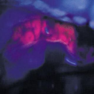
In this video case study, Dr. Hadjipanayis used Gliolan to perform fluorescence guided surgery on a patient with a malignant brain tumor. The surgery was performed using a Leica M530 OH6 neurosurgical microscope with the FL400* intraoperative fluorescence system:
* For all fluorescence modules, please check the status of regulatory approval for your country with your local Leica Microsystems representative.
Contact DB Surgical for more information.
Republished from Leica Microsystems.
